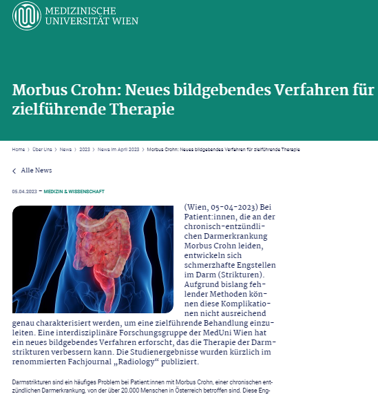
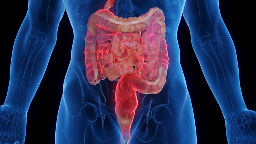
Crohn’s disease is an inflammatory bowel disease that affects the digestive system. It causes inflammation of the lining of the digestive tract, leading to abdominal pain, severe diarrhea, fatigue, weight loss, and malnutrition. In some cases, Crohn’s disease can lead to life-threatening complications such as intestinal obstruction or fistulas.
As part of the interdisciplinary research at the Medical University of Vienna at the University Clinic for Radiology and Nuclear Medicine, a newly developed nuclear medicine tracer was used for precise imaging procedures for the first time. This specific FAPI tracer has the unique property of binding to the Fibroblast-Activating Protein (FAP) present in connective tissue cells, which, in the case of the disease, cause fibrosis of the intestinal wall.
Using PET-MRI, correlations between molecular imaging and the pathological extent of fibrosis were demonstrated. This enabled differentiation between moderate and severe fibrosis grades—a factor crucial for determining the most appropriate treatments.
"With the molecular imaging we have developed, those patients who could benefit from surgical intervention could be identified early on in the future. This would spare them from a less effective drug therapy for fibrotic strictures."
Dr. Michael Bergmann
Evaluation of Intestinal Fibrosis with 68Ga-FAPI PET/MR Enterography in Crohn Disease
Martina Scharitzer, Andrea Macher-Beer, Thomas Mang, Lukas W. Unger, Alexander Haug, Walter Reinisch, Michael Weber, Thomas Nakuz, Lukas Nics, Marcus Hacker, Michael Bergmann, Sazan Rasul
https://doi.org/10.1148/radiol.222389


